All Scanning Services

We cover a wide variety of scans

Neuro Endocrine Tumors

About PET/CT Scanning

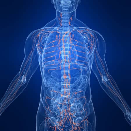
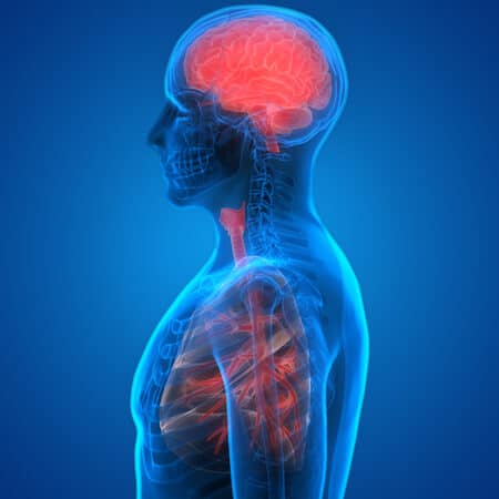
A PET/CT scan combines a PET scan and a CT scan (also known as a CAT scan) into one comprehensive examination unlike any other available today. PET is very good at determining whether or not a tumor is malignant by evaluating its metabolic activity. Most tumors demonstrate an elevated level of activity that functionally distinguishes it from normal tissues. CT scans provide cross-sectional anatomic images (called slices) of your body. Together, these images are called PET/CT, and the power of these two techniques together provides far greater value than either provided alone.

The PET/CT scan is a powerful tool used by doctors when evaluating patients who may or may not have cancer. It allows them to effectively diagnose and evaluate cancer and whether or not it has spread to other areas of the body. It is often used to measure the response of a tumor to therapy, which ultimately guides therapeutic decisions. If a therapy is found to be effective, it can be continued with greater confidence. If found ineffective, more appropriate alternate therapies can be instituted earlier, and futile therapeutic options can be avoided sooner. PET-CT is also used to direct or guide biopsies and surgeries as well as focusing radiation treatments. Once in remission, patients can be reliably monitored for possible recurrence at regular intervals as well.

We also offer pre-authorization services for our referring offices.

PET/CT Lymphoma Scan

Lymphoma

Lymphoma is a cancer of the lymphatic system, which plays an important role in the body’s immune system. In the United States, the risk of developing Non-Hodgkin lymphoma is 2.1 percent and getting Hodgkin Lymphoma is approximately 0.2 percent, and survival depends on a timely diagnosis so that treatment can be given. PET/CT scans are performed when a doctor believes that lymphoma may be present.

How PET Scans Work to Find Lymphoma

PET scans tend to be most useful in those who have Hodgkin lymphoma or high-grade non-Hodgkin lymphoma because the lymphoma cells are typically more active. The PET scan will help the doctor determine which cells are cancerous and which ones aren’t. A PET scan will be combined with a CT scan, which provides accurate images of the body’s organs, lymph nodes, tumors.

When PET Scans are Used

PET scans are typically performed at the beginning or end of a treatment cycle. Before treatment begins, a PET scan is commonly used to determine what stage the lymphoma is and which parts of the body are affected. After treatment, diagnostic imaging will be performed to:

  • Determine the effectiveness of the treatment.
  • Create a future treatment plan based on the results.
  • Determine if the lymphoma has returned a few months or years after treatment.

PET/CT Brain Scan

Using PET/CT to Detect Alzheimer’s and Dementia

PET/CT scans work by using a “tracer” to detect during the scan. Each kind of scan uses a different tracer depending on what the radiologist is trying to find. For brain scans, the tracer is called Amyvid. This tracer was developed recently and approved by the FDA in 2012. It binds to those beta-amyloid proteins, the cause of brain plaque leading to brain cell death. The PET/CT scan picks up the tracker and it allows for early detection of plaque build up.

What Causes Degenerative Brain Disease?

Diseases like Alzheimer’s are caused by continual brain cell death. A connection scientists have made is a connection between brain cell death and plaque buildup. These plaques are clusters of built up proteins called “beta-amyloid”. This kills the brain cell, and from within the dead brain cell, “tangles” are formed. These are fibers of a protein called “tau”. Normally, tau helps transport nutrients between brain cells, but in the presence of tangles, they do just the opposite.

Who would be a candidate for PET/CT brain scans?

According to an article published in 2014, appropriate candidates for amyloid PET imaging evaluation would include:

  1. Patients complaining of persistent or progressive unexplained mild cognitive impairment (MCI)
  2. Patients meeting tests for possible AD but who have unclear clinical presentation, either atypical clinical course or etiologically mixed presentations
  3. Patients with progressive dementia and atypically early age of onset (defined as before the age of 65 years)

Additionally, inappropriate candidates for amyloid PET imaging include

  1. Patients who are 65 years or older and meet standard clinical criteria for probable AD
  2. Those whose severity of disease has to be determined
  3. Those undergoing imaging solely based on a family history of dementia or presence of other risk factors for AD, such as the ApoE-ε4 gene
  4. Patients with a cognitive complaint but no clinical confirmation of impairment
  5. Those undergoing imaging in lieu of genotyping for suspected autosomal mutation carriers
  6. Asymptomatic individuals
  7. Those undergoing imaging for nonmedical use, such as insurance coverage and legal or employment screening.

NETSPOT

NETSPOT, the new tracer for Gallium 68-Dotatate scan, is the next generation of OCTREOSCAN.

It is a form of octreotide, which binds to a receptor on many NETs called the Somatostatin Receptor.  Use of NETSPOT changed patient treatment and management in 71% of patients due to greater specificity compared to Octreoscan.

Gallium PET/CT is more accurate for staging and superior to Octreoscan SPECT in the detection of overall number of lesions in the body as well as organs and bones. Gallium PET/CT also allows for calculation of standardized uptake value, has less whole body radiation, and is performed in less time versus Octreoscan.

A novel PET/CT tracer for the detection of Neuro Endocrine Tumors (NETs)

What makes NETSPOT different?

NETSPOT™ offers superior sensitivity and accuracy compared to previously available diagnostics for NET patients. In a clinical study, NETSPOT™ demonstrated the ability to change management decisions regarding patients in 71% of cases previously imaged with Octreoscan. Saving just one patient from futile surgery can improve the treatment experience overall and significantly reduce the cost of care by reducing repetitive, often frustrating, and limitless diagnostics.

NETSPOT™ is a significant improvement over existing diagnostic tools and will enable us to more accurately stage and better manage patients.

NETSPOT™ is a novel patented kit developed by AAA for the preparation of gallium Ga 68 dotatate for injection, for localization of somatostatin receptor positive neuroendocrine tumors (NETs) in adult and pediatric patients using Positron Emission Tomography (“PET“).

This product has been designated as an orphan drug by the EMA and the FDA.

Please contact PET Imaging Institute of South Florida at 954-981-6668 for further information.

A novel PET/CT tracer for the detection of Neuro Endocrine Tumors (NETs)

NETSPOT, the new tracer for Gallium 68-Dotatate scan, is the next generation of OCTREOSCAN.

It is a form of octreotide, which binds to a receptor on many NETs called the Somatostatin Receptor.  Use of NETSPOT changed patient treatment and management in 71% of patients due to greater specificity compared to Octreoscan.

Gallium PET/CT is more accurate for staging and superior to Octreoscan SPECT in the detection of overall number of lesions in the body as well as organs and bones. Gallium PET/CT also allows for calculation of standardized uptake value, has less whole body radiation, and is performed in less time versus Octreoscan.

Ready To Make An Appointment?

Call Now (954) 981-6668 and Receive Top Quality Service For You or Your Patient

The owner of this website has made a commitment to accessibility and inclusion, please report any problems that you encounter using the contact form on this website. This site uses the WP ADA Compliance Check plugin to enhance accessibility.