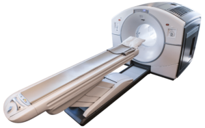
PET Scanning For Prostate Cancer

PET Scanning For Prostate Cancer
If you or a loved one has been diagnosed with any type of cancer, there are often many questions left unanswered. For those diagnosed with prostate cancer, a doctor may suggest having a PET scan as a part of an overall plan for treatment and monitoring for any recurrence or progression of disease. We hope this article may be a resource and help answer a few of the most common questions that we receive regarding PET scanning for prostate cancer.
With prostate cancer being the second leading cause of cancer death in American men (1), new advances in early detection and high-quality imaging for staging and restaging of prostate cancer have become a top priority for the medical community. Comprehensive cancer care requires high-quality imaging and is the driving force behind rapid and ongoing advancements in imaging techniques used for prostate cancer. In recent years, PET scanning for prostate cancer has become an increasingly popular choice to clearly locate and assess the extent of prostate cancer. While there have been many improvements across many conventional imaging modalities such as TRUS, CT, and MRI, this article is intended to highlight the essential role of advanced imaging such as PET scanning for prostate cancer.
Are you looking to learn more about PSMA PET scanning for prostate cancer? Click Here.
If you are in need of a PSMA PET Scan please join our priority list by Clicking Here.
What Is A PET Scan and Why Is It Used for Prostate Cancer?
- What is a PET scan? A “PET Scan”, short for Positron Emission Tomography Scan, is an imaging technique that uses radioactive tracers to clearly image targeted areas in the body. It is primarily used in the diagnosis, initial staging and treatment strategy, to assess the effectiveness of therapy and for restaging, or the evaluation for recurrent disease after treatment.
- Why is a PET scan used for prostate cancer? PET scanning is used for prostate cancer because of its superior ability to target and capture images of prostate cancer on a cellular level. This allows for more accurate staging and restaging in the overall prostate cancer treatment strategy. With new radiotracers being developed and studied, PET scanning continues to lead the way in what will be possible for imaging during treatment of prostate cancer.
PET scanning has revolutionized the way prostate cancer is imaged because of its ability to target cancer on a cellular and molecular level. PET Scanning uses a radioactive tracer that is absorbed and visualized in cancerous cells related to the prostate. This makes a PET scan a better choice than more conventional imaging modalities that only evaluate an anatomical “snapshot” of abnormalities rather than the functional aspects of a PET scan.
A PET scan is combined with a CT scan, acquired at the same time, which is why you may sometimes read or hear someone refer to this procedure as a PET/CT scan. With CT capturing high-quality anatomical images and PET detecting changes on the molecular level, the combination of these allows for the creation of extremely accurate superimposed single images that no other imaging modality can replicate. This level of precision and accuracy is why PET scanning is often the number one choice when trying to determine the clearest path forward for many patients with prostate cancer. Specifically, at this time, this is predominantly utilized in men with suspected recurrence of prostate cancer after treatment. A PET scan can detect and characterize very small areas of recurrent prostate cancer, even with PSA levels that are very low.
Additional PET Scanning FAQ
Is PET scanning safe?
While PET scanning does involve the use of radioactive tracers, these are “diagnostic levels” of radiation that are completely safe and have no known side effects.
How long does it take to get the results after a PET Scan?
Images are captured and created during the PET scan procedure, and afterward, a radiologist will utilize their training and experience to interpret the scan and produce a written report of findings and conclusions. This report is then transmitted to your referring physician who would then review those results with you and discuss any further treatment if needed.
What is it like for a patient to have a PET Scan?
Please watch this video we created that clearly walks one through the experience of having a PET scan and answers all common questions about PET scanning.
When and Why are PET Scans used for Prostate Cancer?
- Staging: A PET Scan is used for patients with known prostate cancer in order to pinpoint its exact location and to determine the extent of disease and to determine a treatment strategy.
- Planning Treatment: In some instances, a PET scan may be used to specifically target certain high-risk areas for special treatment. There are occasions when a PET scan is the only diagnostic test that can identify these otherwise hidden areas of cancer spread.
- Evaluation During and After Treatment: A PET Scan can be used during and after treatment of prostate cancer to determine the effectiveness or response of specific drugs and therapies.
- Ongoing Cancer Care After Detection of Biochemically Recurrent Prostate Cancer – Restaging: A PET scan allows physicians to locate and assess the extent of recurrent prostate cancer. Specifically, a new radiopharmaceutical called Axumin (see below) has a superior ability to detect recurrent prostate cancer, even in very early stages and in patients with low PSA levels.
General Example of PET Scan Use After A Prostate Cancer Diagnosis
It is important to note that while PET Scanning can be an essential tool in the assessment of prostate cancer, it is not always used on all patients, and there are many other imaging tests and procedures that may be recommended depending on the patient’s specific needs.
This is an example of a prostate cancer care that would include PET scanning.
- Prostate cancer is detected by the results of screening PSA (prostate-specific antigen) with a blood test or a DRE (digital rectal exam).
- Prostate cancer is confirmed by a core needle biopsy with imaging (TRUS or MRI) either before or during the procedure.
- In some instances, a PET scan may be used during the initial evaluation and treatment strategy. Prostate cancer is evaluated after a diagnosis using various imaging techniques to determine the extent or stage of cancer. While imaging modalities like MRIs are often used to stage prostate cancer, PET scanning has been used effectively to stage prostate cancer and new tests like PSMA PET scans continue to show promise in being the best future solutions during prostate cancer staging.
- A PET scan is usually the most effective way to restage recurrent prostate cancer. After prostate cancer treatment, PET scanning may be used to determine the effectiveness of the treatment and is now commonly being used for the detection of biochemically recurrent prostate cancer for restaging. A specific PET radioactive tracer, Axumin, has been designed for this exact purpose of targeting and restaging recurrent prostate cancer.
What Are the Most Common “Types” of PET Scans For Prostate Cancer?
- PSMA (Prostate-specific Membrane Antigen) PET Scan: Now FDA approved, they are used to locate, stage, and restage prostate cancer. PSMA PET scans have been FDA approved to be used after a diagnosis of prostate cancer in order to stage and determine if cancer has spread to other parts of the body. They have also been approved for use in locating and restaging recurrent prostate cancer for patients with biochemical recurrence.
- Axumin (Fluciclovine F-18) PET Scan: Currently in use primarily for the purpose of restaging recurrent prostate cancer in those patients with suspected biochemical recurrence.
- FDG (Fluorodeoxyglucose) PET Scan: May also be used for staging in particular cases of prostate cancer. In some instances, depending on the prostate cancer tumor type, FDG PET scans may be used for the restaging of recurrent prostate cancer as well.
PSMA (Prostate-Specific Membrane Antigen) PET Scanning For Prostate Cancer Care
PSMA PET scans are now FDA-approved and ongoing studies continue to suggest that it will be an even more valuable imaging tool used for both staging and restaging prostate cancer in the near future. The specifics of PSMA will hopefully allow for even more accurate imaging and the earliest possible detection when performing PET scans for prostate cancer.
When Is A PSMA PET Scan Used For Prostate Cancer?
Now FDA approved, we are currently performing PSMA PET Scans at our facility for the following purposes.
- Staging: Patients with a first-time diagnosis of prostate cancer benefit from using a PSMA PET scan because of its superior ability to detect cancer spread — offering the best possible future treatment options.
- Restaging: Patients with suspected biochemically recurrent prostate cancer, meaning patients with elevated PSA levels after definitive therapy, benefit from using PSMA PET scans because of its ability to precisely locate recurrent prostate cancer and detect cancer spread. For both of these reasons, PSMA PET scans offer the best treatment options for recurrent prostate cancer.
How Do PSMA (Prostate-Specific Membrane Antigen) PET Scans Work?
PSMA PET scans use a different functional method of imaging prostate cancer cells. PSMA is a glycoprotein, a type of sugar-protein that resides on the surface of prostate cancer cells. These tracers, such as PSMA, are injected and then PET scan images are acquired in the standard manner. As with all PET scans, these images are then interpreted by a radiologist and subsequently used to guide treatment strategy.
Axumin (Fluciclovine F-18) PET Scanning For Prostate Cancer Care
Axumin is an FDA-approved agent used for Axumin PET scans for prostate cancer. Axumin is often able to image and restage recurrent prostate cancer better than any other conventional imaging techniques. Biochemical recurrence, typically suspected with rising PSA (prostate-specific antigen) levels, is the standard in monitoring patients for suspected recurrent prostate cancer. Traditional imaging techniques are often limited in that they may detect a small lymph node or ‘suspicious’ finding, but cannot further functionally characterize the molecular activity to determine the level of suspicion. The introduction of Axumin PET scanning has been a breakthrough, allowing physicians the ability to accurately locate and restage prostate cancer with precision, especially in the setting of suspiciously rising PSA levels.
How Do Axumin (Fluciclovine F-18) PET Scans Work?
An Axumin PET uses a radioactive tracer, given as an injection, that is linked to an amino acid which is absorbed by prostate cancer at a much more rapid rate than normal cells. The rapid uptake of Axumin by prostate cancer cells is then imaged by the advanced technology within the PET scan equipment. The PET scan images are then reviewed in order to determine if there has been any spread to other areas in the body.
Next Steps
PET Imaging Institute is a leading provider in PET scanning and has over 20 years of experience in using PET scans for prostate cancer. We understand that you may still have questions about why your doctor has suggested a PET scan for you or someone in your family.
If your doctor has requested a PET scan for prostate cancer as part of your medical care, you may have come across this article as part of a search for more information. We believe in keeping our patients informed and up-to-date on the latest advances in PET scans, and PET scans for prostate cancer making those advancements available to you. Whether you are a local, international, or someone who frequents South Florida in need of these services, please email us at psma@piisf.com to discuss your next steps and have us answer any questions you may have!
Already Have A PSMA PET Scan Order From Your Doctor?
Send your prescription to: psma@piisf.com or use our PSMA Fax Hotline: (954) 607-6757
Schedule your PSMA PET scan appointment by clicking here.
References


